Tackling Cancer with Tech
Thu, 04/09/2020 - 12:00
Better diagnostic tools, coupled with Artificial Intelligence-assisted analytics and data sharing platforms, are helping clinicians improve medical outcomes for cancer patients.
Hippocrates—whose oath modern physicians still hold sacred—ascribed cancer to ‘an excess of black bile’ in various parts of the body. Bound by the knowledge and tools of his time, he was inadvertently wrong.
Nonetheless, it was he who named cancer, christening it with the Greek word for ‘crab’. The tumours he saw—solid lumps with protrusions of blood vessels at their periphery—reminded him of the hard-shelled crustaceans, and perhaps he was intentionally alluding to their difficult-to-access nature.
Fast forward to the present day, and the scourge of cancer remains. In fact, the World Health Organisation reports that the number of cancer cases is on the rise globally, and one in six individuals will die of the disease.
But the situation is less dire than it seems; scientists and clinicians are adding new ‘weapons’ to their arsenal of anticancer drugs, while others are devising more sensitive and accurate ways to catch the disease in its early stages, when the chances of survival are much higher. Throw Artificial Intelligence (AI) and surgical robotics into the mix and physicians today are increasingly empowered to treat and extend the lives of cancer patients.
Insight from the Inside
At the very beginning of every cancer patient’s journey is a diagnosis, without which any subsequent treatment may be misdirected and ineffective. It comes as no surprise, then, that new diagnostic tools are constantly being developed and validated to detect cancer.
Since many cancers are nestled deep inside the body, sprouting from traitorous, fast-growing cells in various tissues and organs, being able to see beneath the body’s surface is essential for doctors to make a preliminary diagnosis. Medical imaging technologies such as ultrasound scans and magnetic resonance imaging may allow doctors to view potentially cancerous lumps inside a patient, but to definitively conclude that the lump is malignant, a needle biopsy is typically performed.
During a needle biopsy, a small piece of biological tissue is excised from the suspected tumour for analysis. Simple as this procedure may seem, it requires a significant amount of skill on the part of the surgeon.
“The clinician or interventional radiologist will have to determine where they insert the needle from, considering factors like the obstruction by the ribs and even blood vessels,” said Mr Alan Goh, Co-founder and CEO of NDR Medical Technology, a Singapore-based firm specialising in surgical robotics. “Get it wrong, and you might cause the collapse of a lung or serious internal bleeding.” A clinician’s experience thus plays a big part in charting the course of the biopsy needle.
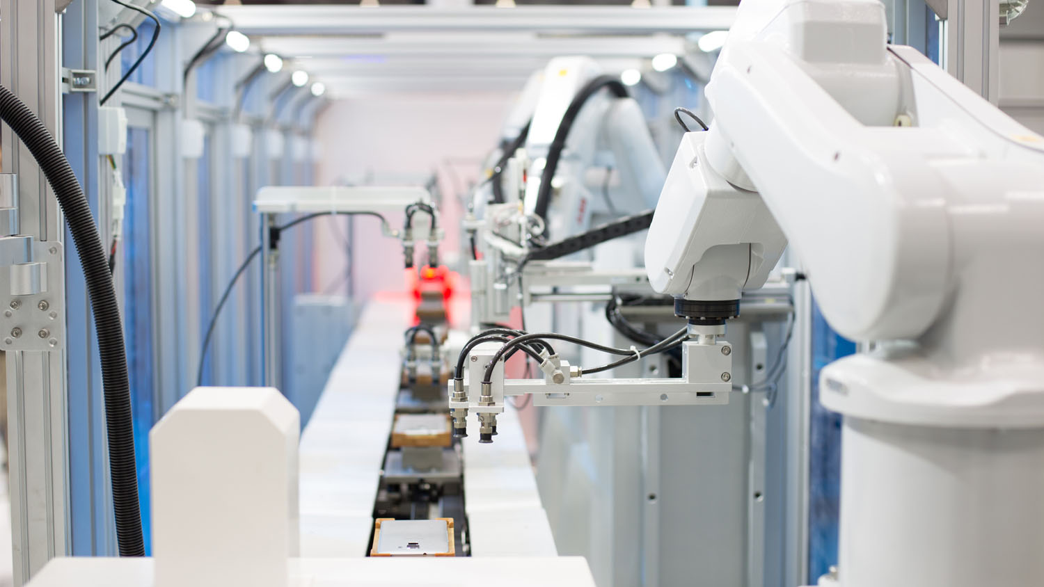
Seeking to enable clinicians of all experience levels to perform biopsies safely and efficiently, Mr Goh has developed the Automated Needle Targeting system. The combination of AI for image analysis and surgical robotics allows for precise planning and execution of biopsies—based on a medical image, the software recommends the best path to access the tumour and aligns the surgical robot accordingly. Subsequently, the clinician is guided by the surgical robot along the recommended path, and out comes the tissue sample.
“It takes only seconds for our software to pinpoint the trajectory for the clinician and align the surgical robot, so the entire biopsy procedure can be completed in less than three minutes,” Mr Goh explained, adding that the system is a skills-leveller, allowing for more consistent and precise biopsies.
An Algorithm for Efficiency
Once a biopsy has been obtained from the patient, it is passed on to the pathology department for further processing. This typically involves slicing the tissue sample into microscopically thin sections, mounting those sections on glass slides, then staining them with dyes that help pathologists distinguish between benign and malignant cells.
Cancer, especially in its later stages, alters the organisation and composition of the tissue it resides in, and this can be spotted in the biopsy sections. In some cases, however, the changes are more subtle, requiring a well-trained eye to spot malignancy. Then there are many biopsies of suspicious lumps that are not cancer at all but get funnelled through the pathology department just to rule them out.
“So pathologists end up having to spend a lot of time looking for things that aren’t there, adding to their substantial workload,” said Mr Dan Hosseinzadeh, CEO and Co-founder of Pathcore, a digital pathology solutions firm based in Toronto, Canada. “What Pathcore aims to do with our technology is to eliminate work that is not clinically meaningful so that pathologists can focus their attention on the things that matter to patients.”
With the PathcoreSedeen slide viewer, for example, pathologists can either use Pathcore’s image analysis tools or even create their analysis algorithms to facilitate and expedite their decision making. Qritive, another digital pathology platform developer based in Singapore, adopts a similar approach, providing its users with tissue-specific AI modules to identify cancer cells among millions of normal cells.
“Our Pantheon AI system analyses microscopy images of the patient’s tissue samples, and the results are shown to the pathologist as annotations on the tissue image. The pathologist then uses these inputs, along with the associated patient history, to arrive at the final diagnosis,” said Qritive’s CEO and Co-founder Dr Aneesh Sathe.
The application of a robust analysis algorithm also reduces inter-observer variability, while the ability to perform quantification of image features raises the confidence of diagnoses. “With quantification, you can get a clearer picture of the stage or grade of cancer,” Mr Hosseinzadeh said.
To further enhance the accuracy of diagnoses, Mr Hosseinzadeh thinks that it is important to break down the silos between different data sources such as pathology slides, medical images or genomic sequences. “Even in silos, there’s so much information we can extract from those datasets, so just imagine what could happen if we break those silos down,” he quipped. Anticipating this ‘explosion and cross-pollination’ of datasets, Pathcore is positioning itself as a broker and integrator of different data types.
Impatient for Better Patient Outcomes
Although biopsies are the current gold standard of cancer diagnosis, clinicians and researchers are reimagining how cancer might be detected in the body. Instead of having to remove tissue from a patient, why not simply draw blood?
This idea has not escaped the notice of Dr Tan Min-Han, CEO and medical director of genomics testing firm Lucence Diagnostics. His company has developed genomic tests that allow clinicians to pick up cancer signals in the blood—what is known as a liquid biopsy. When cancer cells die, they shed DNA fragments into the blood. Lucence’s technology analyses these DNA fragments to identify specific mutations in each patient’s tumour.
“As you can imagine, a liquid biopsy is quicker, and the costs are lower, especially considering all the procedural costs of taking a tissue biopsy and its downstream analysis,” Dr Tan said, adding that a blood test is essentially non-invasive, which reduces patients’ pain and lowers the risk of complications associated with tissue biopsies.
“At Lucence, we depend on Deep Learning approaches for the best medical test results,” Dr Tan said, noting that his analytics team integrates information from a wide range of sources—about the patient, the stage of the disease and the treatments that have been given or are available—to help clinicians make better treatment decisions for cancer patients.
The convergence of digital technology and medicine is already yielding results in clinics and hospitals. “In our work with frontline oncologists, we certainly are seeing patients with lung, colon and breast cancer receive the right treatments after following our tests, and when we follow up with these patients, we see a positive response to the selected treatment regiments,” he said.
At the same time, digital pathology is shrinking the time gap between when a diagnostic test is ordered, and when the results are delivered. “What used to be a two-week waiting period for results from a tissue biopsy can now be completed within 48 hours,” said Mr Yusuke Aoi, vice president of innovation and growth at Parkway Pantai Ltd.
Clearly, a strong synergy exists between the medical community and MedTech companies like Lucence Diagnostics, NDR Medical Technologies, Qritive and Pathcore, and this model of collaboration looks set to spawn further innovation. “Clinicians are not averse to using advanced technologies,” said Dr Sathe. “Doctors ask the most pertinent questions and have a very good understanding of new technologies.”
Patients are the ultimate beneficiaries of this synergistic relationship. “For patients, new medical technology and innovation translates to faster turnaround times and more effective, personalised treatments,” Mr Aoi concluded.
At SGInnovate, we are committed to helping entrepreneurial scientists scale their companies for the betterment of the world. MedTech is one of the areas that we are investing in as we believe in the importance and impact of technology in the healthcare sector.
Qritive, NDR Medical Technologies and Lucence Diagnostics are our portfolio companies.
Trending Posts
- A Guide to Singapore’s Cell & Gene Therapy Ecosystem
- A Guide to Singapore’s Hydrogen Ecosystem
- Walking the tightrope of disclosure to create a robust IP strategy
- Why intellectual property (IP) strategy can mean the difference between life and death for a startup
- Going behind-the-scenes in a MedTech startup for a 6-month internship to create lasting impact